ミクロ相分離構造を有する高分子材料表面 に粘着した血小板形態の電顕的解析
Transmission and scanning of rat platelets copolymer adhered surface with electron to HEMA-St microphase microscopic analysis ABA type block separated structure
Kazuhiko ABE*, Kazunori KATAOKA*, Morie SEKIGUCHI**,
Teruo OKANO***, Yasuhisa SAKURAI***, Isao SHINOHARA****
and Akira SHIMADA****
Polymeric material which has microphase separated structure is a most promising candidate for antithrombogenic material.
In order to investigate the role of microphase separated structure on the interaction between polymers and platelets, HEMA-St ABA type block copolymers with microphase separated structure which is composed of hydrophilic 2-hydroxyethyl methacrylate
(HEMA) and hydrophobic styrene (St) were prepared. By chang
ing mole fraction of HEMA in copolymers, block copolymers with the following three types of microphase separated structure were obtained : (1) isolated hydrophilic islands in continuous hydrophobic phase (sea-island microstructure), lamellar microstructure
(2) which is composed of alternative hydrophilic and hydrophobic phases, (3) isolated hydrophobic islands in continuous hydrophilic phasereversed) sea-island microstructure).
Rat platelets suspended in Hanks’ balanced salt solution (Ca,
Mg++ free) were used for the experiment. Interaction of platelets with polymer surface was studied by microsphere columnar method. The morphological changes of adhering platelets on polymer surfaces were investigated by transmission and scanning
electron microscopy.
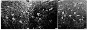
Platelets adhered to the surface of lamellar microstructure have smooth surface with round shape and short pseudopods, indicating that the morphological changes are less marked than those of platelets adhered to the surfaces of both sea-island and reversed sea-island microstructure. Although platelets attached to the surfaces of the three types of microphase separated structures retained organella such as a-granules and mitochondria relatively well, extended open canalicular system was observed in platelets adhered to the surfaces of sea-island and reversed sea-island microstructure.
These findings suggest that the surface of lamellar microstructure
has an inhibitory action on platelet activation as compared with the surfaces of sea-island and reversed sea-island microstructure.
Thus, the mode of platelet adhesion was found to be greatly influenced by the domain shape and size of microphase separated structure.
It is concluded that the morphology of the microphase separated structure is an important element for the molecular design of excellent antithrombogenic material.

※What an amazing team! When I was young, I saw a lot of platelets!

